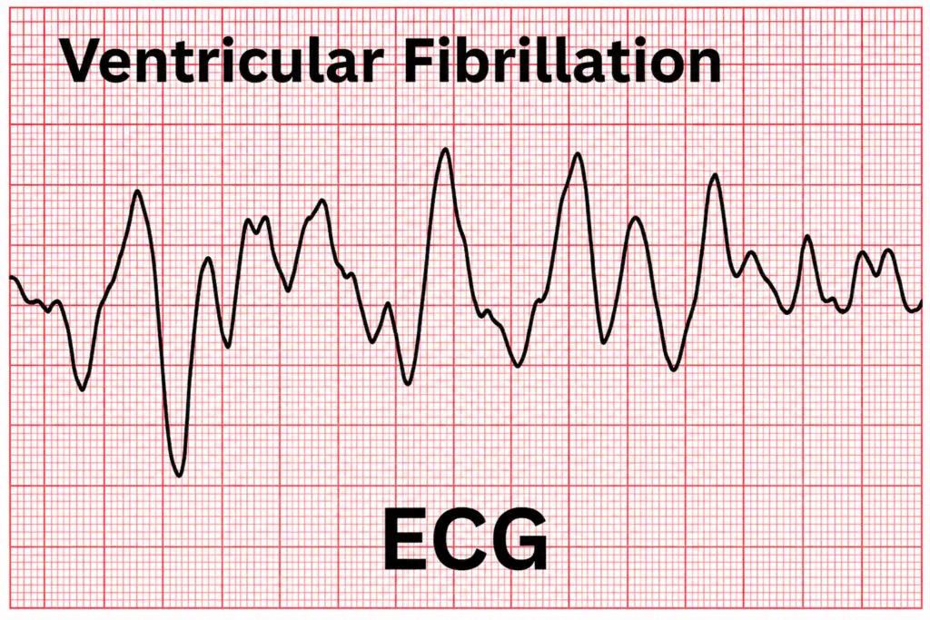
Ventricular Fibrillation and Cardiac Arrest: Not the Same Thing
Written by Stephanie Austin, Owner & Lead Trainer, Prima Cura Training |Last reviewed: August 2026 | Next review: August 2027
In the previous post in this series, I looked at the difference between a heart attack and cardiac arrest. Two terms that most people use interchangeably, and shouldn’t. This post goes one layer deeper.
If you’ve ever watched an AED demonstration or been through CPR training, you’ll have heard the phrase “shockable rhythm.” The device analyses the heart, announces its verdict, and either instructs you to deliver a shock or tells you none is advised. But what is it actually detecting? And where does ventricular fibrillation fit in?
The short answer: ventricular fibrillation is the most common cause of sudden cardiac arrest, and it’s the rhythm AEDs are primarily designed to treat. But VF and cardiac arrest are not the same thing. Understanding the distinction gives you a much clearer picture of what you’re actually doing when you put an AED on someone’s chest — and why it sometimes tells you no shock is needed.
At a Glance: VF vs Cardiac Arrest
| Ventricular Fibrillation (VF) | Cardiac Arrest | |
| What it is | A specific abnormal heart rhythm, A chaotic, rapid electrical activity in the ventricles | The state of the heart is not pumping blood effectively. It is an outcome, not a rhythm |
| Same thing? | No, VF is a cause | No, cardiac arrest is the result |
| Shockable? | Yes | Only if caused by a shockable rhythm (VF or pulseless VT) |
| AED helps? | Yes | Only if the underlying rhythm is shockable |
| Only cause? | No, but the most common one | Can result from VF, pulseless VT, PEA, or asystole |
What Is Ventricular Fibrillation?
The heart’s pumping action is controlled by electrical signals. In a healthy heart, those signals fire in a coordinated sequence: the upper chambers (the atria) contract first, pushing blood down into the lower chambers (the ventricles), which then contract to push blood out to the lungs and the rest of the body. That coordinated cycle, repeated sixty to one hundred times per minute, is what keeps you alive.
Ventricular fibrillation is what happens when that electrical system breaks down completely. Instead of a single, organised signal triggering each contraction, the ventricles receive a storm of rapid, chaotic electrical impulses. Instead of contracting and pumping, they quiver. The ventricles may be firing two hundred times a minute or more, but none of those impulses produce a useful contraction. No blood is being pumped. No oxygen is reaching the brain.
On an ECG, VF appears as a fast, erratic waveform with no discernible pattern whatsoever. Nothing resembling the neat, regular peaks and troughs of a normal sinus rhythm.
VF can come on with no warning. It can be triggered by a heart attack, by severe electrolyte imbalances, by a significant electric shock, by certain medications, or by inherited abnormalities in the heart’s electrical system – conditions such as long QT syndrome or Brugada syndrome. In some cases, particularly in sudden cardiac death in otherwise healthy adults and young people, VF occurs in a structurally normal heart with no obvious prior cause. It is one of the reasons why defibrillators in schools, sports facilities, and public spaces are not optional extras.
What Is Cardiac Arrest?
Cardiac arrest is a state, not a rhythm. It’s what happens when the heart stops pumping blood effectively, regardless of what’s causing that failure. The person becomes unresponsive, stops breathing normally, and will die without immediate intervention.
I covered cardiac arrest in detail in the previous post, including the signs to look for and exactly what to do. If you haven’t read that one yet, it’s worth starting there. For the purposes of this post, the key point is this: cardiac arrest is the outcome. VF is one of the causes.
So VF and Cardiac Arrest Are Not the Same Thing?
Correct. VF is one of several cardiac rhythms that can result in cardiac arrest. When VF causes cardiac arrest, the heart is in that specific chaotic electrical state. But cardiac arrest can also be caused by other rhythms, some of which look very different and require a different clinical response.
The distinction matters most when we talk about shockable and non-shockable rhythms. VF is shockable. Not all cardiac arrest rhythms are. Understanding is what turns “follow the AED’s instructions” from a rote instruction into something you genuinely understand.
The Four Rhythms of Cardiac Arrest
When someone is in cardiac arrest, their heart is in one of four electrical states. Two of them respond to defibrillation. Two of them don’t.
| Rhythm | What it means | Shockable? | AED response |
| Ventricular fibrillation (VF) | Rapid, chaotic electrical activity. Ventricles quiver instead of pump. | Yes | Shock advised |
| Pulseless ventricular tachycardia (pVT) | Fast, organised but ineffective rhythm. No pulse. Same outcome as VF. | Yes | Shock advised |
| Pulseless electrical activity (PEA) | Organised electrical signals present but no effective mechanical pumping. | No | No shock, CPR only |
| Asystole | No discernible electrical activity. End-stage or presenting rhythm. | No | No shock, CPR only |
Ventricular fibrillation (VF)
The most common cause of sudden cardiac arrest is the one with the best survival outlook if treated quickly. The ventricles are receiving chaotic electrical signals and quivering instead of pumping. Defibrillation can reset the rhythm by simultaneously depolarising the heart muscle cells, giving the heart’s own natural pacemaker the opportunity to reassert control. The AED will recommend a shock.
Pulseless ventricular tachycardia (pulseless VT)
VT is a fast but organised abnormal rhythm originating in the ventricles. When it’s sufficiently rapid and the person has no detectable pulse, the outcome is functionally the same as VF: no effective pumping, no circulation. It is a shockable rhythm. The AED will recommend a shock.
Pulseless electrical activity (PEA)
This is one of the most counterintuitive cardiac arrest states. The heart’s electrical system is producing recognisable signals. An ECG would show organised activity. But the mechanical pumping function has failed. The electrical signal is present; the contraction is not. PEA can be caused by severe blood loss, a tension pneumothorax, cardiac tamponade, massive pulmonary embolism, or severe metabolic disturbance, among others. Defibrillation cannot help here. The electrical system isn’t the problem. The AED will not recommend a shock, and this is correct. CPR maintains circulation while paramedics work to identify and treat the underlying cause.
Asystole
Often called “flatlining,” though the dramatic straight-line monitor beep of television medicine is a reasonable creative liberty rather than a clinical description. Asystole means there is no discernible electrical activity in the heart at all. It can be an end-stage rhythm following prolonged, untreated VF, or it can present as the first rhythm in certain types of cardiac arrest. Like PEA, it is non-shockable. A defibrillation shock will not restart a heart in asystole. The AED will not recommend a shock. CPR is the priority.
What Does “Shockable” Actually Mean?
A shockable rhythm is one that can potentially be corrected by a controlled electrical shock delivered through the chest wall to the heart muscle. The logic behind it is this: in VF and pulseless VT, the heart is producing disorganised or abnormal electrical activity. A defibrillation shock delivers a large, brief burst of electrical energy, which simultaneously depolarises the myocardial cells. It doesn’t so much restart the heart as it resets it, briefly stopping all electrical activity at once and creating the conditions for the sinoatrial node, the heart’s natural pacemaker, to reassert a normal rhythm.
It doesn’t always work on the first shock. That’s why current resuscitation protocols involve resuming CPR immediately after a shock rather than waiting to see if the rhythm has corrected. Continued CPR maintains coronary perfusion pressure during those critical seconds, which in turn improves the likelihood that subsequent shocks will be effective.
For PEA and asystole, there is no chaotic electrical activity to reset. The problem is either mechanical (PEA) or a complete absence of electrical activity (asystole). Delivering a shock to a heart in either state will not help and will delay the CPR that actually can.
What Does the AED Actually Do?
Three things.
It analyses the heart rhythm. The pads placed on the chest act as electrodes, reading the electrical activity. The AED’s algorithm assesses whether the rhythm falls into a shockable category. This analysis takes around ten seconds, which is why you’re told to stop CPR and stand clear during that window — movement creates electrical interference that can confuse the reading.
If the rhythm is shockable, it charges and instructs you to deliver a shock. The shock itself lasts milliseconds. Everyone must stand completely clear. The current delivered is strong enough to cause injury to anyone in contact with the patient.
It tells you what to do next. Modern AEDs provide continuous voice guidance, prompting CPR after the shock and telling you when to allow it to re-analyse. Follow the instructions exactly. The AED is not infallible, but it is considerably more accurate at rhythm analysis than any bystander.
| Important: What the AED does not do The AED does not restart a stopped heart. This is one of the most persistent misconceptions about defibrillation, probably reinforced by the dramatic “clear!” paddles scenes in every medical drama ever made. The AED corrects a chaotic or abnormal rhythm in a heart that is still producing electrical activity. If the shock is successful and normal rhythm is restored, the heart starts pumping again. But the AED’s role is rhythm correction, not resuscitation in isolation. CPR is what keeps oxygenated blood reaching the brain in the minutes before and after. Neither intervention alone is as effective as both together. |
What Does This Mean for a First Aider in Practice?
When you’re responding to a cardiac arrest, you won’t know which rhythm the person is in. You can’t tell VF from asystole without an ECG, and you’re not expected to. Your job is the same regardless:
- Start CPR immediately
- Get the AED on as fast as possible
- Follow the AED’s instructions
- Keep CPR going, including after a shock
The AED handles the diagnosis. What understanding the underlying rhythms does for you is something more valuable than that: it means you understand why the AED sometimes says “no shock advised.” Not because something has gone wrong. Not because the person is beyond help. But because the rhythm they’re in doesn’t respond to defibrillation, CPR is the most important thing you can do for them right now.
That understanding makes a real difference to how confidently someone acts in a genuine emergency. It’s the reason I take the time to explain the mechanics in every BLS and AED course I run, rather than just drilling the steps. Informed first aiders respond better. The evidence supports it.
The Resuscitation Council UK is clear that early CPR and early defibrillation are the two interventions most likely to improve survival from out-of-hospital cardiac arrest. Both matter. Neither should wait.
Train With Someone Who Explains the Why
Understanding what an AED is actually doing and why we do what we do isn’t just interesting. It changes how confidently you respond when it counts. The steps are the same for everyone. The difference is in whether you’re following instructions or whether you genuinely understand them.
Every course I run covers both theory and hands-on practice, because one without the other only gets you so far.
Basic Life Support and AED | Emergency First Aid at Work (EFAW) | First Aid at Work (FAW)
Get in touch: info@primacuratraining.co.uk | 0333 999 8783
Frequently Asked Questions
Is ventricular fibrillation the same as cardiac arrest?
No. VF is a specific abnormal heart rhythm characterised by rapid, chaotic electrical activity in the ventricles that produces no effective pumping. Cardiac arrest is the state of the heart not circulating blood effectively. VF is one of the most common causes of sudden cardiac arrest, but cardiac arrest can also result from pulseless VT, pulseless electrical activity (PEA), and asystole.
What are the four rhythms of cardiac arrest?
The four rhythms associated with cardiac arrest are ventricular fibrillation (VF), pulseless ventricular tachycardia (pulseless VT), pulseless electrical activity (PEA), and asystole. VF and pulseless VT are shockable: an AED may be able to correct them. PEA and asystole are non-shockable: CPR and treatment of the underlying cause are the priority, and a shock will not help.
Why does an AED sometimes say “no shock advised”?
Because the heart is in a non-shockable rhythm, if the AED detects PEA or asystole, delivering a shock cannot help. There is no disorganised electrical activity to reset. The correct response is to continue CPR. If the AED says no shock is advised, it is working exactly as it should. Keep doing CPR and do not delay questioning the device.
Can VF be treated without a defibrillator?
No. CPR alone cannot correct VF, but it is essential while a defibrillator is being retrieved. CPR maintains circulation to the brain and vital organs, buying critical time until the AED arrives. Without CPR, the window for successful defibrillation closes rapidly. Without defibrillation, VF cannot be corrected. Both are needed, and neither should be delayed.
What causes ventricular fibrillation in an otherwise healthy person?
VF can occur in structurally normal hearts due to inherited electrical abnormalities such as long QT syndrome, Brugada syndrome, or catecholaminergic polymorphic ventricular tachycardia (CPVT). These conditions can be present with no prior symptoms. This is the mechanism behind many cases of sudden cardiac death in young people and athletes, and it is one of the strongest arguments for accessible defibrillators in schools and sports facilities.
This article is intended for general information purposes only and does not constitute medical or professional advice. All information reflects current Resuscitation Council UK guidance and is correct as of August 2026. Medical guidance is subject to change; always refer to current official sources for up-to-date information. In an emergency, call 999 immediately.